

Time Bound Regression of Pituitary Hyperplasia in Primary Pituitary Hypothyroidism Following Treatment
Article Sidebar
Views | PDF/EPUB Downloads:
219
/ 31
/ 18
Main Article Content
Abstract
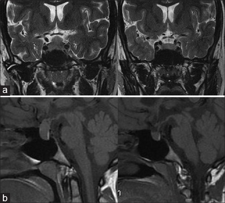
Aim: The aim was to prospectively study radiological regression of pituitary hyperplasia following the treatment for primary
hypothyroidism by serial magnetic resonance imaging (MRI).
Subjects and Methods: Seven patients (1 male and 6 females) with profound hypothyroidism were included, age ranging from 13 to 42 years. MRI for pituitary was done at first visit and on follow‑up. Pituitary size on imaging was matched with baseline T3, T4, thyroid-stimulating hormone (TSH), and thyroid peroxidase antibody antibodies and TSH on follow-up.
Results: By 6 weeks all patients showed clinical improvement with near normalization of thyroid function tests in most patients. The TSH normalization was well correlated with regression of the pituitary enlargement on imaging at 6 weeks. The height of pituitary gland at baseline was 13.24 ± 3.61 mm, at first follow‑up 9.68 ± 1.32 mm, and at second follow‑up 7.80 ± 3.95 mm. The diameter of the pituitary gland at baseline was 14.47 ± 4.14 mm, at first follow‑up 11.83 ± 3.99 mm, and at second follow‑up 7.25 ± 4.59 mm. The height of the pituitary gland significantly reduced in first and second follow‑up (P < 0.05) and reduction in diameter on second follow-up (P < 0.05).
Conclusion: Pituitary hyperplasia secondary to primary hypothyroidism regresses with treatment within 6 weeks of initiation of treatment in its height however, it takes months for reduction in the diameter of the gland. We recommend repeat imaging in enlarged and homogenously enhancing pituitary gland in patients of primary hypothyroidism at 6 weeks of initiation of treatment to prove or disprove hyperplasia.
Downloads
Article Details
Section

This work is licensed under a Creative Commons Attribution-NonCommercial 4.0 International License.
This is an open access journal, and articles are distributed under the terms of the Creative Commons Attribution-NonCommercial-ShareAlike 4.0 License, which allows others to remix, tweak, and build upon the work non-commercially, as long as appropriate credit is given and the new creations are licensed under the identical terms.
How to Cite
References
1. Babu S, Venkataramana NK, Kamble RB, Rao SA, Naik A, Shetty RV. Primary hypothyroidism presenting as a sellar mass which regressed with thyroid hormone therapy. A case report. Neuroradiol J 2010;23:433‑6.
2. Xu AJ, Li T. Pituitary hyperplasia secondary to primary hypothyroidism in children: Report of 8 cases. Zhongguo Dang Dai Er Ke Za Zhi 2010;12:17‑20.
3. Shimono T, Hatabu H, Kasagi K, Miki Y, Nishizawa S, Misaki T, et al. Rapid progression of pituitary hyperplasia in humans with primary
hypothyroidism: Demonstration with MR imaging. Radiology 1999;213:383‑8.
4. Franceschi R, Rozzanigo U, Failo R, Bellizzi M, Di Palma A. Pituitary hyperplasia secondary to acquired hypothyroidism: Case report. Ital J Pediatr 2011;37:15.
5. Simsek E, Simsek T, Savas‑Erdeve S, Erdogmus B, Dösoglu M. Pituitary hyperplasia mimicking pituitary macroadenoma in two adolescent patients with long‑standing primary hypothyroidism: Case reports and review of literature. Turk J Pediatr 2009;51:624‑30.
6. Young M, Kattner K, Gupta K. Pituitary hyperplasia resulting from primary hypothyroidism mimicking macroadenomas. Br J Neurosurg 1999;13:138‑42.
7. Yamada T, Tsukui T, Ikejiri K, Yukimura Y, Kotani M. Volume of sella turcica in normal subjects and in patients with primary hypothyroidism and hyperthyroidism. J Clin Endocrinol Metab 1976;42:817‑22.
8. Sarlis NJ, Brucker‑Davis F, Doppman JL, Skarulis MC. MRI‑demonstrable regression of a pituitary mass in a case of primary hypothyroidism after a week of acute thyroid hormone therapy. J Clin Endocrinol Metab 1997;82:808‑11.
9. Hutchins WW, Crues JV 3rd, Miya P, Pojunas KW. MR demonstration of pituitary hyperplasia and regression after therapy for hypothyroidism. AJNR Am J Neuroradiol 1990;11:410.