

Chest and upper abdominal computed tomography scan findings in patients with established breast cancer
Article Sidebar
Views | PDF/EPUB Downloads:
738
/ 147
/ 56
Main Article Content
Abstract
Background: Computed tomography (CT) of the chest and/or abdomen is usually done in patients with breast cancer for staging regardless of their grade, size, lymph node, or clinical signs/symptoms in Nigeria. This study aimed to determine the diagnostic yield of chest and upper abdominal CT for metastasis, metastatic pattern, and incidental findings in patients with breast cancer in our environment.
Methodology: A retrospective study of all 300 confirmed breast cancer patients who reported for chest/abdominal computed tomographic scans in a tertiary diagnostic center in Lagos between October 2021 and December 2021. Data were extracted from their CT images/radiological reports and analyzed using SPSS version 23 for Windows, P <0.05.
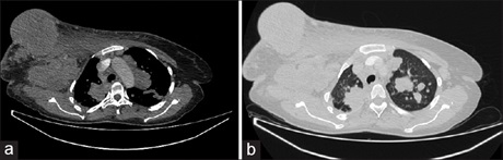
Results: Three hundred patients with established breast cancer were examined, within the age range of 24–83 years, median of 51 years, and mean of 50.91 ± 11.9 years. The majority were female 298 (99.3%) and within the 50–59-year (101, 33.7%) age group. Most had unilateral breast cancer 286 (95%) with left-sided predominance (146, 49%). Metastasis was seen in 183 (61%) patients with nodes being the most common site 158 (52.7%). Solitary metastasis was seen in 91 (30.3%) of the participants, predominating in the lymph nodes 69 (23%), followed by lungs 17 (5.7%), P < 0.01. Two-organ involvement was in 59 (19.7%), and the most common combination was lung and node 32 (10.7%), followed by liver and node 8 (2.7%). Metastasis was most common in the 50–59-year age group, 64 (21.3%).
Conclusion: Chest and abdominal CT yielded a definitive diagnosis of metastasis in more than 50% of the patients. The most common structure affected was the lymph nodes. Chest CT is therefore imperative in patients with late presentation of breast cancer.
Downloads
Article Details
Section
This is an open access journal, and articles are distributed under the terms of the Creative Commons Attribution-NonCommercial-ShareAlike 4.0 License, which allows others to remix, tweak, and build upon the work non-commercially, as long as appropriate credit is given and the new creations are licensed under the identical terms.
How to Cite
References
1. Jedy-Agba EE, Dos-Santos-Silva I, Olaomi O, Yilkudi M, Ezeome E, Idris S. Delays in breast cancer presentation and diagnosis in Nigeria.
J Clin Oncol 2016;34:15 Suppl: e13092. Available from: https://www.ascopubs.org/doi/abs/10.1200/JCO.2016.34.15_suppl.e13092. [Last
accessed on 2020 Apr 24].
2. Barrett T, Bowden DJ, Greenberg DC, Brown CH, Wishart GC, Britton PD. Radiological staging in breast cancer: Which asymptomatic
patients to image and how. Br J Cancer 2009;101:1522-8.
3. Ravaioli A, Pasini G, Polselli A, Papi M, Tassinari D, Arcangeli V, et al. Staging of breast cancer: New recommended standard procedure.
Breast Cancer Res Treat 2002;72:53-60.
4. Ciatto S, Pacini P, Azzini V, Neri A, Jannini A, Gosso P, et al. Preoperative staging of primary breast cancer. A multicentric study.Cancer 1988;61:1038-40.
5. Crivello ML, Ruth K, Sigurdson ER, Egleston BL, Evers K, Wong YN, et al. Advanced imaging modalities in early stage breast cancer:
Preoperative use in the United States Medicare population. Ann Sur Oncol 2013;20:102-10.
6. Kim H, Han W, Moon HG, Min J, Ahn SK, Kim TY, et al. The value of preoperative staging chest computed tomography to detect
asymptomatic lung and liver metastasis in patients with primary breast carcinoma. Breast Cancer Res Treat 2011;126:637-41.
7. Gerber B, Seitz E, Müller H, Krause A, Reimer T, Kundt G, et al. Perioperative screening for metastatic disease is not indicated in
patients with primary breast cancer and no clinical signs of tumor spread. Breast Cancer Res Treat 2003;82:29-37.
8. James J, Teo M, Ramachandran V, Law M, Stoney D, Cheng M. Acritical review of the chest CT scans performed to detect asymptomatic
synchronous metastasis in new and recurrent breast cancers. World J Surg Oncol 2019;17:40.
9. Dull B, Linkugel A, Margenthaler JA, Cyr AE. Overuse of chest CT in patients with stage I and II breast cancer: An opportunity to increase
guidelines compliance at an NCCN member institution. J Natl Compr Canc Netw 2017;15:783-9.
10. Ahn SJ, Kim YS, Kim EY, Park HK, Cho EK, Kim YK, et al. The value of chest CT for prediction of breast tumor size: Comparison
with pathology measurement. World J Surg Oncol 2013;11:130
11. Son JH, Jung HK, Song JW, Baek HJ, Doo KW, Kim W, et al. Incidentally detected breast lesions on chest CT with US correlation:
A pictorial essay. Diagn Interv Radiol 2016;22:514-8.
12. Smith-Bindman R. Environmental causes of breast cancer and radiation from medical imaging: Findings from the institute of
medicine report. Arch Intern Med 2012;172:1023-7.
13. Omidiji OA, Campbell PC, Irurhe NK, Atalabi OM, Toyobo OO. Breast cancer screening in a resource poor country: Ultrasound versus
mammography. Ghana Med J 2017;51:6-12.
14. Olubunmi Obajimi M, Adeniji-Sofoluwe AT, Adeoye AO, Obajimi GO, Ajani MA, Adejumo PO, et al. Bilateral breast cancer among three
Yoruba women in a Nigerian teaching hospital. BJR Case Rep 2015;1:16.
15. Sharma D, Singh G. Breast cancer in young women: A retrospective study from tertiary care center of North India. South Asian J Cancer
2017;6:51-3.
16. Perkins CI, Hotes J, Kohler BA, Howe HL. Association between breast cancer laterality and tumor location, United States, 1994-1998. Cancer Causes Control 2004;15:637-45.
17. Weiss HA, Devesa SS, Brinton LA. Laterality of breast cancer in the United States. Cancer Causes Control 1996;7:539-43.
18. Roychoudhuri R, Putcha V, Møller H. Cancer and laterality: A study of the five major paired organs (UK). Cancer Causes Control
2006;17:655-62.
19. Tulinius H, Sigvaldason H, Olafsdóttir G. Left and right sided breast cancer. Pathol Res Pract 1990;186:92-4.
20. Sughrue T, Brody JP. Breast tumor laterality in the United States depends upon the country of birth, but not race. PLoS One 2014;9:e103313.
21. Chen MT, Sun HF, Zhao Y, Fu WY, Yang LP, Gao SP, et al. Comparison of patterns and prognosis among distant metastatic breast cancer patients by age groups: A SEER population-based analysis. Sci Rep 2017;7:9254.
22. James J, Teo M, Ramachandran V, Law M, Stoney D, Cheng M. Performance of CT scan of abdomen and pelvis in detecting
asymptomatic synchronous metastasis in breast cancer. Int J Surg 2017;46:164-9.
23. Berman AT, Thukral AD, Hwang WT, Solin LJ, Vapiwala N. Incidence and patterns of distant metastases for patients with early-stage
breast cancer after breast conservation treatment. Clin Breast Cancer 2013;13:88-94.
24. Patanaphan V, Salazar OM, Risco R. Breast cancer: Metastatic patterns and their prognosis. South Med J 1988;81:1109-12.
25. Kutomi G, Ohmura T, Satomi F, Takamaru T, Shima H, Suzuki Y, et al. Lymph node shape in computed tomography imaging as a predictor for axillary lymph node metastasis in patients with breast cancer. Exp Ther Med 2014;8:681-5.
26. Skok K, Hladnik G, Grm A, Crnjac A. Malignant pleural effusion and its current management: A review. Medicina (Kaunas) 2019;55:490.
27. Incidental Findings. American College of Radiology. Available from https://www.acr.org/clinical-resources/incidental-findings. [Last
accessed on 2020 Apr 24].
28. Mehta LS, Watson KE, Barac A, Beckie TM, Bittner V, Cruz-Flores S, et al. Cardiovascular disease and breast cancer: Where these entities
intersect: A scientific statement from the American Heart Association. Circulation 2018;137:e30-66.