

sonographic Evaluation of Gallbladder Dimensions in Healthy Adults in Benin City, Nigeria
Article Sidebar
Views | PDF/EPUB Downloads:
340
/ 61
/ 51
Main Article Content
Abstract
Background: Several disease conditions can affect gallbladder (GB) size and wall thickness (WT). Imaging methods are superior to
clinical evaluation in assessing GB dimensions. Ultrasonography is a relatively safe, inexpensive and reproducible imaging modality for
assessing normal or diseased GB. There are few reports on normal GB dimensions in the Nigerian medical literature. This study therefore set out to contribute to data on GB dimensions among Nigerians.
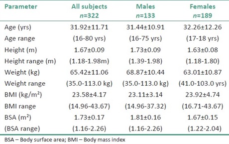
Materials and Methods: This was a prospective study. Three hundred and twenty-two healthy adult volunteers, consisting of 133 males and 189 females were assessed, by ultrasound, following over night fasting. GB length, width, height and WT were measured for each subject. GB-V was calculated by the ellipsoid formula. Data analysis included descriptive statistics and comparison of measurements with biometric parameters. Statistical significance between the variables was done with the Students t-test, with ‘P’ value set at ≤0.05.
Results: One hundred and thirty-three males (41.3%) and one hundred and eighty-nine females (58.7%) were studied. The mean age of subjects was 31.92±11.7 years. The mean values of the length (L), height (H), and width (W) of the GB were 6.16±1.09 cm; 2.75±0.58 cm; and 2.98±0.59 cm respectively. Mean GB-V was 27.2±12.8 cm3 and WT 0.25±0.04 cm. Age and gender did not significantly influence GB measurements.
Conclusions: A normal range of GB dimensions for the Benin City locality has been established. The study confirmed the non-dependence of GB measurements on age and gender.
Downloads
Article Details
Section

This work is licensed under a Creative Commons Attribution-NonCommercial 4.0 International License.
This is an open access journal, and articles are distributed under the terms of the Creative Commons Attribution-NonCommercial-ShareAlike 4.0 License, which allows others to remix, tweak, and build upon the work non-commercially, as long as appropriate credit is given and the new creations are licensed under the identical terms.
How to Cite
References
1. Berman MC. Gallbladder and biliary system. In: Kwamura DM, editor. Diagnostic Medical Sonography: A Guide to Clinical Practice, Abdomen and Superficial Structures. 2nd ed. Philadelphia: Lippincott‑Raven; 1997. p. 191‑235.
2. Karani J. The abdomen. In: Sutton D, editor. Textbook of Radiology and Imaging. 7 th ed., Vol. 1. Philadelphia: Churchill Livingstone; 2003. p. 711‑8.
3. Berthold B. The Practice of Ultrasound: A Step to Step Guide to Abdominal Scanning. Stuttgart: George Thieme Verlag; 2004. p. 106‑39.
4. Ugwu AC. Body surface area as a surrogate measure of gallbladder sizes and indices; a predictive equation in humans. Sudan JMS 2007; 2:101‑4.
5. Olokoba B, Bojuwoye BJ, Olokoba LB, Wahab KW, Braimoh KT, Inikori AK, et al. The relationship between gallstone disease and gallbladder wall thickness. Afr Sci 2006;7:171‑6.
6. Sanders RC, Winter T. Clinical Sonography: A Practical Guide. 4 th ed. Philadelphia: Lippincott Williams and Wilkins; 2007. p. 81‑93.
7. Dodds WJ, Groh WJ, Darweesh RM, Lawson TL, Kishk SM, Kern MK. Sonographic measurement of gallbladder volume. AJR Am J Roentgenol 1985;145:1009‑11.
8. Caroli‑Bosc FX, Pugliese P, Peten EP, Demarquay JF, Montet JC, Hastier P, et al. Gallbladder volume in adults and its relationship to age, sex, body mass index, body surface area and gallstones. An epidemiologic study in a nonselected population in France. Digestion 1999;60:344‑8.
9. Nieves MA, Bueno J, Gaona Yánez C, Mercedes González M. Comparative study of gallbladder volume and contraction in healthy subjects of various ages and sex by ultrasonography. GEN 1989;43:13‑7.
10. Sari R, Balci MK, Coban E, Karayalcin U. Sonographic evaluation of gallbladder volume and ejection fraction in obese women without gallstones. J Clin Ultrasound 2003;31:352‑7.
11. Braverman DZ, Johnson ML, Kern F Jr. Effects of pregnancy and contraceptive steroids on gallbladder function. N Engl J Med 1980; 302: 362‑4.
12. Agarwal AK, Miglani S, Singla S, Garg U, Dudeja RK, Goel A. Ultrasonographic evaluation of gallbladder volume in diabetics. J Assoc Physicians India 2004;52:962‑5.
13. Kishk SM, Darweesh RM, Dodds WJ, Lawson TL, Stewart ET, Kern MK, et al. Sonographic evaluation of resting gallbladder volume and postprandial emptying in patients with gallstones. AJR Am J Roentgenol 1987;148:875‑9.
14. Ngige EN, Renner JK, Temiye EO, Njokanma OF, Arogundade RA, David AN. Ultrasonographic measurement of the hepatobiliary axis of children with sickle cell anaemia in steady state. Niger J Clin Biomed Res 2006;1:44‑50.
15. Yoo JH, Kwak HJ, Lee MJ, Suh JS, Rhee CS. Sonographic measurements of normal gallbladder sizes in children. J Clin Ultrasound 2003;31:80‑4.
16. Gourtsoyiannis NC, Damilakis JE, Charoulakis NZ , Bakantaki AS, Vlahonikolis JG, Xynos E. Relationship of gallbladder contour, fasting volume and emptying to body size indices in normal subjects and patients with gallstones. Digestion 1995;56:395‑9.