

Should Susceptibility‑weighted Imaging be Included in the Protocol for Evaluation of Acute Ischemic Stroke Patients?
Article Sidebar
Views | PDF/EPUB Downloads:
17
/ 2
/ 1
Main Article Content
Abstract
Context: Stroke is one of the leading causes of death globally and is a major cause of long‑term disability.
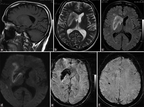
Aims of Study: (1) To identify hemorrhagic foci in patients with acute ischemic stroke by susceptibility‑weighted imaging (SWI). (2) To compare the detection of hemorrhagic foci in patients with acute ischemic stroke by SWI versus conventional magnetic resonance imaging (MRI) (T1‑weighted, T2‑weighted, and fluid‑attenuated inversion recovery [FLAIR]).
Materials and Methods: Two‑hundred and fifty patients, who clinically presented with neurological deficit, were evaluated using 1.5 Tesla MRI scanner from October 2011 to September 2013 with the above sequences. Detection of hemorrhage in acute infarct was evaluated and compared.
Results: Of 250 patients evaluated in this study, 232 cases were arterial infarcts and 18 cases were venous infarcts. Hemorrhage was detected in 86 (34.4%) patients, of which 68 cases were from arterial infarcts and 18 were from venous infarcts. SWI was significantly sensitive and specific (P < 0.004) for the detection of hemorrhage in acute infarct compared to T1‑weighted, T2‑weighted, and FLAIR.
Conclusion: SWI is a very sensitive sequence for the detection of hemorrhage in acute stroke patients. Therefore, its use is recommended in the protocol for evaluation of these patients.
Downloads
Article Details
Section

This work is licensed under a Creative Commons Attribution-NonCommercial 4.0 International License.
This is an open access journal, and articles are distributed under the terms of the Creative Commons Attribution-NonCommercial-ShareAlike 4.0 License, which allows others to remix, tweak, and build upon the work non-commercially, as long as appropriate credit is given and the new creations are licensed under the identical terms.
How to Cite
References
1. Garcia JH, Mitchem HL, Briggs L, Morawetz R, Hudetz AG, Hazelrig JB, et al. Transient focal ischemia in subhuman primates. Neuronal injury as a function of local cerebral blood flow. J Neuropathol Exp Neurol 1983;42:44‑60.
2. Bell BA, Symon L, Branston NM. CBF and time thresholds for the formation of ischemic cerebral edema, and effect of reperfusion in baboons. J Neurosurg 1985;62:31‑41.
3. Hossmann KA, Schuier FJ. Experimental brain infarcts in cats. I. Pathophysiological observations. Stroke 1980;11:583‑92.
4. Schuier FJ, Hossmann KA. Experimental brain infarcts in cats. II. Ischemic brain edema. Stroke 1980;11:593‑601.
5. Heiss WD, Rosner G. Functional recovery of cortical neurons as related to degree and duration of ischemia. Ann Neurol 1983; 14:294‑301.
6. Nighoghossian N, Hermier M, Adeleine P, Blanc‑Lasserre K, Derex L, Honnorat J, et al. Old microbleeds are a potential risk factor for cerebral bleeding after ischemic stroke: A gradient‑echo T2*‑weighted brain MRI study. Stroke 2002;33:735‑42.
7. Reichenbach JR, Venkatesan R, Schillinger DJ, Kido DK, Haacke EM. Small vessels in the human brain: MR venography with deoxyhemoglobin as an intrinsic contrast agent. Radiology 1997;204:272‑7.
8. Mittal S, Wu Z, Neelavalli J, Haacke EM. Susceptibility‑weighted imaging: Technical aspects and clinical applications, part 2. AJNR
Am J Neuroradiol 2009;30:232‑52.
9. Santhosh K, Kesavadas C, Thomas B, Gupta AK, Thamburaj K, Kapilamoorthy TR. Susceptibility weighted imaging: A new tool
in magnetic resonance imaging of stroke. Clin Radiol 2009;64:74‑83.
10. Weingarten K, Zimmerman RD, Deo‑Narine V, Markisz J, Cahill PT, Deck MD. MR imaging of acute intracranial hemorrhage:
Findings on sequential spin‑echo and gradient‑echo images in a dog model. AJNR Am J Neuroradiol 1991;12:457‑67.
11. Atlas SW, Thulborn KR. MR detection of hyperacute parenchymal hemorrhage of the brain. AJNR Am J Neuroradiol 1998; 19:1471‑7.
12. Linfante I, Llinas RH, Caplan LR, Warach S. MRI features of intracerebral hemorrhage within 2 hours from symptom onset. Stroke 1999;30:2263‑7.
13. Liang L, Korogi Y, Sugahara T, Shigematsu Y, Okuda T, Ikushima I, et al. Detection of intracranial hemorrhage with susceptibility‑weighted MR sequences. AJNR Am J Neuroradiol 1999;20:1527‑34.
14. Kidwell CS, Saver JL, Villablanca JP, Duckwiler G, Fredieu A, Gough K, et al. Magnetic resonance imaging detection of microbleeds before thrombolysis: An emerging application. Stroke 2002;33:95‑8.
15. Hermier M, Nighoghossian N, Derex L, Berthezène Y, Blanc‑Lasserre K, Trouillas P, et al. MRI of acute post‑ischemic cerebral hemorrhage in stroke patients: Diagnosis with T2*‑weighted gradient‑echo sequences. Neuroradiology 2001;43:809‑15.