Role of Preoperative Multidetector Computed Tomography Diagnosis of Solid Pseudopapillary Tumors of the Pancreas with Postoperative Surgical and Histopathological Correlation
Main Article Content
Abstract
Purpose: The purpose of this study was to evaluate the role of preoperative multidetector computed tomography (MDCT)
diagnosis of solid pseudopapillary tumors (SPTs) of the pancreas with postoperative surgical and histopathological correlation.
Materials and Methods: A prospective study was conducted in our institute for MDCT evaluation of patients with ultrasound‑proven pancreatic tumors. Preoperative diagnosis of SPT was given in 10 of 36 total patients evaluated. These findings were correlated with surgical and histopathological findings.
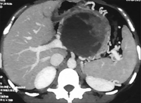
Results: A preoperative MDCT diagnosis of SPT was given in 10 patients on the basis of characteristic CT appearances, of which 9 were confirmed by postoperative histopathology. One was a histopathological examination (HPE) proven to be a neuroendocrine tumor. Two MDCT‑negative but HPE‑positive cases gave a total of 11 of 36 patients. 10 patients were females with a mean age of 27 (range 16–38 years). 6 lesions were identified in the head, with the average size of
the lesions being 6.5 cm. No SPTs with malignant features were diagnosed on MDCT or HPE in our study. The sensitivity of MDCT to identify SPT in this series is 81.81%, specificity 96%, positive predictive value of 90%, negative predictive value of 92.31%.
Conclusion: MDCT has a high specificity and positive predictive value with higher negative predictive values for diagnosing SPTs. However, atypical lesions pose a diagnostic challenge. A diagnosis with a greater degree of confidence can be made using knowledge of characteristic appearance on MDCT along with clinical correlation. The majority are benign, but follow‑up is suggested if signs of aggressiveness are identified radiologically or by HPE.
Downloads
Article Details
Section

This work is licensed under a Creative Commons Attribution-NonCommercial 4.0 International License.
This is an open access journal, and articles are distributed under the terms of the Creative Commons Attribution-NonCommercial-ShareAlike 4.0 License, which allows others to remix, tweak, and build upon the work non-commercially, as long as appropriate credit is given and the new creations are licensed under the identical terms.
How to Cite
References
1. Trivedi N, Sharma U, Das PM, Mittal MK, Talib VH. FNAC of papillary and solid epithelial neoplasm of pancreas –A case report.
Indian J Pathol Microbiol 1999;42:369‑72.
2. Stömmer P, Kraus J, Stolte M, Giedl J. Solid and cystic pancreatic tumors. Clinical, histochemical, and electron microscopic features
in ten cases. Cancer 1991;67:1635‑41.
3. Procacci C, Graziani R, Bicego E, Zicari M, Bergamo Andreis IA, Zamboni G, et al. Papillary cystic neoplasm of the pancreas:
Radiological findings. Abdom Imaging 1996;21:554‑8.
4. Hamilton SR, Altonen LA. World Health Organization Classification of Tumours. Tumours of the Digestive System. Lyon: IARC Press; 2000. p. 246‑8. Available from: http://www.w2.iarc.fr/en/publications/pdfs‑online/pat‑gen/bb2/BB2.pdf.
5. Barreto G, Shukla PJ, Ramadwar M, Arya S, Shrikhande SV. Cystic tumours of the pancreas. HPB (Oxford) 2007;9:259‑66.
6. Crawford BE 2nd. Solid and papillary epithelial neoplasm of the pancreas, diagnosis by cytology. South Med J 1998;91:973‑7.
7. Patil TB, Shrikhande SV, Kanhere HA, Saoji RR, Ramadwar MR, Shukla PJ. Solid pseudopapillary neoplasm of the pancreas: A single institution experience of 14 cases. HPB (Oxford) 2006;8:148‑50.
8. Lam KY, Lo CY, Fan ST. Pancreatic solid‑cystic‑papillary tumor: Clinicopathologic features in eight patients from Hong Kong and
review of the literature. World J Surg 1999;23:1045‑50.
9. Jung SE, Kim DY, Park KW, Lee SC, Jang JJ, Kim WK. Solid and papillary epithelial neoplasm of the pancreas in children. World J Surg 1999;23:233‑6.
10. Choi BI, Kim KW, Han MC, Kim YI, Kim CW. Solid and papillary epithelial neoplasms of the pancreas: CT findings. Radiology 1988; 166:413‑6.
11. Huang HL, Shih SC, Chang WH, Wang TE, Chen MJ, Chan YJ. Solid‑pseudopapillary tumor of the pancreas: Clinical experience and literature review. World J Gastroenterol 2005;11:1403‑9.
12. Mao C, Guvendi M, Domenico DR, Kim K, Thomford NR, Howard JM. Papillary cystic and solid tumors of the pancreas: A pancreatic embryonic tumor? Studies of three cases and cumulative review of the world’s literature. Surgery 1995;118:821‑8.
13. Podevin J, Triau S, Mirallié E, Le Borgne J. Solid‑pseudopapillary tumor of the pancreas: A clinical study of five cases, and review
of the literature. Ann Chir 2003;128:543‑8.
14. Coleman KM, Doherty MC, Bigler SA. Solid‑pseudopapillary tumor of the pancreas. Radiographics 2003;23:1644‑8.
15. Wang DB, Wang QB, Chai WM, Chen KM, Deng XX. Imaging features of solid pseudopapillary tumor of the pancreas on
multi‑detector row computed tomography. World J Gastroenterol 2009;15:829‑35.
16. Choi JY, Kim MJ, Kim JH, Kim SH, Lim JS, Oh YT, et al. Solid pseudopapillary tumor of the pancreas: Typical and atypical
manifestations. AJR Am J Roentgenol 2006;187:W178‑86.
17. Buetow PC, Buck JL, Pantongrag‑Brown L, Beck KG, Ros PR, Adair CF. Solid and papillary epithelial neoplasm of the pancreas: Imaging‑pathologic correlation on 56 cases. Radiology 1996;199:707‑11.
18. Cantisani V, Mortele KJ, LevyA, Glickman JN, Ricci P, PassarielloR, et al. MR imaging features of solid pseudopapillary tumor of the
pancreas in adult and pediatric patients. AJR Am J Roentgenol 2003;181:395‑401.
19. YuMH, Lee JY, KimMA, Kim SH, Lee JM, Han JK, et al. MR imaging features of small solid pseudopapillary tumors: Retrospective
differentiation from other small solid pancreatic tumors. AJR Am J Roentgenol 2010;195:1324‑32.
20. Sunkara S, Williams TR, Myers DT, Kryvenko ON. Solid pseudopapillary tumours of the pancreas: Spectrum of imaging findings with histopathological correlation. Br J Radiol 2012;85:e1140‑4.


