3‑Tesla Magnetic Resonance Imaging evaluation of intraspinal extramedullary nonosseous neoplastic lesions with their postsurgical and histopathological correlation
Main Article Content
Abstract
Aims: This study aimed to evaluate the diagnostic ability of 3-Tesla magnetic resonance imaging (MRI) in detection and characterization of intraspinal extramedullary nonosseous neoplastic lesions and their histopathological correlation.
Settings and Design: It was a prospective study involving fifty patients who presented with low backache and lower limb weakness with MRI findings suggestive of a neoplastic extramedullary intraspinal lesion.
Patients and Methods: A 3-Tesla 16-channel MRI scanner was used for imaging. Multiplanar imaging was done using T1, T2, short-tau inversion recovery sequences and T1 fat-saturated sequences. All patients then underwent postgadolinium MRI. The findings of MRI were then reviewed. An analysis of correlation between MRI findings and surgical and histopathological findings was done.
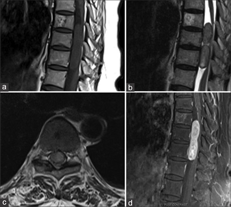
Results: Out of the fifty patients evaluated, intradural lesions were noted in forty patients and extradural lesions in ten patients. Schwannoma was the most common tumor, followed by meningioma in the intradural category. Two rare cases of intradural lipoma and malignant peripheral nerve sheath tumors were also detected. In extradural category, metastasis was the most common lesion along with two rare lesions of epidural meningioma and leukemia-associated granulocytic sarcoma.
Conclusions: MRI is an indispensable tool in the evaluation of spinal neoplasms. Its multiplanar capability, high-quality soft-tissue resolution, and depiction of various anatomic landmarks help the neurosurgeon to make a road map for the surgery. In most of the cases, MRI can give information regarding the histopathology of the lesion. However, in some cases, differential diagnosis needs to be included, especially in cases of extradural meningiomas and malignant peripheral nerve sheath tumors, because of their rare incidence.
Downloads
Article Details
Section

This work is licensed under a Creative Commons Attribution-NonCommercial-ShareAlike 4.0 International License.
This is an open access journal, and articles are distributed under the terms of the Creative Commons Attribution-NonCommercial-ShareAlike 4.0 License, which allows others to remix, tweak, and build upon the work non-commercially, as long as appropriate credit is given and the new creations are licensed under the identical terms.
How to Cite
References
1. Porchet F, Sajadi A, Villemure JG. Spinal tumors: Clinical aspects, classification and surgical treatment. Schweiz Rundsch Med Prax 2003; 92:1897‑905.
2. Hufana V, Tan JS, Tan KK. Microsurgical treatment for spinal tumours. Singapore Med J 2005;46:74‑7.
3. Albanese V, Platania N. Spinal intradural extramedullary tumors. Personal experience. J Neurosurg Sci 2002;46:18‑24.
4. Carra BJ, Sherman PM. Intradural spinal neoplasms: A case based review. J Am Osteopath Coll Radiol 2013;2:13‑21.
5. Kocher B, Smirniotopoulos JG, Smith AB. Intradural spinal lesions. Appl Radiol 2009;9.
6. McCormick PC, Post KD, Stein BM. Intradural extramedullary tumors in adults. Neurosurg Clin N Am 1990;1:591‑608.
7. Intradural, Extramedullar y, Schwannoma. Available from: http://www.diagnosticimaging.com/case‑studies/intradural‑extramedullary‑schwannoma#sthash.rMtLryUh.dpuf. [Last accessed on 2017 Apr 12].
8. Ross JS, Moore KR, Shah LM, Borg B, Crim J. Neoplasms, cysts, and other masses: Neoplasms. Diagnostic Imaging Spine. 2nd ed. Manitoba,
Canada: Amirsys; 2010. p. 84-87.
9. Frank BL, Harrop JS, Hanna A, Ratliff J. Cervical extradural meningioma: Case report and literature review. J Spinal Cord Med 2008;31:302‑5.
10. Salehpour F, Zeinali A, Vahedi P, Halimi M. A rare case of intramedullary cervical spinal cord meningioma and review of the literature. Spinal Cord 2008;46:648‑50.
11. Sahni D, Harrop JS, Kalfas IH, Vaccaro AR, Weingarten D. Exophytic intramedullary meningioma of the cervical spinal cord. J Clin Neurosci
2008;15:1176‑9.
12. Arnautovic K, Arnautovic A. Extramedullary intradural spinal tumors: A review of modern diagnostic and treatment options and a report
of a series. Bosn J Basic Med Sci 2009;9 Suppl 1:40‑5.
13. Saydam O, Senol O, Schaaij‑Visser TB, Pham TV, Piersma SR, Stemmer‑Rachamimov AO, et al. Comparative protein profiling reveals minichromosome maintenance (MCM) proteins as novel potential tumor markers for meningiomas. J Proteome Res 2010;9:485‑94.
14. Solero CL, Fornari M, Giombini S, Lasio G, Oliveri G, Cimino C, et al. Spinal meningiomas: Review of 174 operated cases. Neurosurgery
1989;25:153‑60.
15. Levy WJ Jr., Bay J, Dohn D. Spinal cord meningioma. J Neurosurg 1982;57:804‑12.
16. Matsumoto S, Hasuo K, Uchino A, Mizushima A, Furukawa T, Matsuura Y, et al. MRI of intradural‑extramedullary spinal neurinomas and meningiomas. Clin Imaging 1993;17:46‑52.
17. Quekel LG, Versteege CW. The “dural tail sign” in MRI of spinal meningiomas. J Comput Assist Tomogr 1995;19:890‑2.
18. Kumar K, Malik S, Schulte PA. Symptomatic spinal arachnoid cysts: Report of two cases with review of the literature. Spine (Phila Pa 1976)
2003;28:E25‑9.
19. Silbergleit R, Brunberg JA, Patel SC, Mehta BA, Aravapalli SR. Imaging of spinal intradural arachnoid cysts: MRI, myelography and CT.
Neuroradiology 1998;40:664‑8.
20. Andrews BT, Weinstein PR, Rosenblum ML, Barbaro NM. Intradural arachnoid cysts of the spinal canal associated with intramedullary cysts.
J Neurosurg 1988;68:544‑9.
21. Khosla A, Wippold FJ 2nd. CT myelography and MR imaging of extramedullary cysts of the spinal canal in adult and pediatric patients.
AJR Am J Roentgenol 2002;178:201‑7.
22. Petridis AK, Doukas A, Barth H, Mehdorn HM. Spinal cord compression caused by idiopathic intradural arachnoid cysts of the spine: Review of the literature and illustrated case. Eur Spine J 2010;19 Suppl 2:S124‑9.
23. Secer HI, Anik I, Celik E, Daneyemez MK, Gonul E. Spinal hydatid cyst mimicking arachnoid cyst on magnetic resonance imaging. J Spinal
Cord Med 2008;31:106‑8.
24. Beall DP, Googe DJ, Emery RL, Thompson DB, Campbell SE, Ly JQ, et al. Extramedullary intradural spinal tumors: A pictorial review. Curr
Probl Diagn Radiol 2007;36:185‑98.
25. Shen SH, Lirng JF, Chang FC, Lee JY, Luo CB, Chen SS, et al. Magnetic resonance imaging appearance of intradural spinal lipoma. Zhonghua
Yi Xue Za Zhi (Taipei) 2001;64:364‑8.
26. Blount JP, Elton S. Spinal lipomas. Neurosurg Focus 2001;10:e3.
27. Levy WJ, Latchaw J, Hahn JF, Sawhny B, Bay J, Dohn DF. Spinal neurofibromatosis: A case report of 66 cases and a comparison with
meningiomas. Neurosurgery 1986;18:331‑4.
28. Murphey MD, Smith WS, Smith SE, Kransdorf MJ, Temple HT. From the archives of the AFIP. Imaging of musculoskeletal neurogenic tumors: Radiologic‑pathologic correlation. Radiographics 1999;19:1253‑80.
29. Pileri SA, Ascani S, Cox MC, Campidelli C, Bacci F, Piccioli M, et al. Myeloid sarcoma: Clinico‑pathologic, phenotypic and cytogenetic analysis of 92 adult patients. Leukemia 2007;21:340‑50.
30. Frohna BJ, Quint DJ. Granulocytic sarcoma (chloroma) causing spinal cord compression. Neuroradiology 1993;35:509‑11.
31. Wasserstrom WR, Glass JP, Posner JB. Diagnosis and treatment of leptomeningeal metastases from solid tumors: Experience with 90 patients. Cancer 1982;49:759‑72.


