

Utility of contrast‑enhanced fluid‑attenuated inversion recovery in magnetic resonance imaging of intracranial lesions
Article Sidebar
Views | PDF/EPUB Downloads:
411
/ 48
/ 34
Main Article Content
Abstract
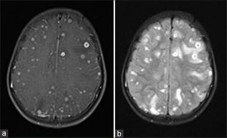
Aim: The aim of this study is to determine utility of contrast-enhanced fluid-attenuated inversion recovery (FLAIR) imaging by comparing results with contrast-enhanced T1-weighted imaging (T1WI) in various intracranial lesions.
Materials and Methods: Forty-nine patients with a known intracranial lesion or with clinical suspicion underwent the gadolinium-enhanced magnetic resonance (MR) imaging using 1.5T. Postcontrast axial, coronal, and sagittal T1 fat-saturated, axial FLAIR images were acquired after administration of gadobenate dimeglumine. The MR imaging parameters for the postcontrast T2-FLAIR images were 6000–9000/90–110/1845–2030 ms/150 (repetition time/echo time/inversion time/flip angle), and the acquisition time was 2 min 12 s. All images were acquired with a section thickness of 5 mm, an intersection gap of 2 mm, and a field of view of 256 mm × 144 mm. The images were transferred to a workstation and reviewed.
Results: We found that postcontrast FLAIR images are useful by showing better meningeal involvement in various pathologies and enhancement of the solid component in intra-axial lesions. However, it was not much helpful in extra-axial lesions and lesions with mild postcontrast enhancement and lesions with perilesional edema.
Conclusion: Postcontrast FLAIR is a useful adjunct to postcontrast T1W images in equivocal cases and for additional information.
Downloads
Article Details
Section

This work is licensed under a Creative Commons Attribution-NonCommercial-ShareAlike 4.0 International License.
This is an open access journal, and articles are distributed under the terms of the Creative Commons Attribution-NonCommercial-ShareAlike 4.0 License, which allows others to remix, tweak, and build upon the work non-commercially, as long as appropriate credit is given and the new creations are licensed under the identical terms.
How to Cite
References
1. Lee EK, Lee EJ, Kim S, Lee YS. Importance of contrast‑enhanced fluid‑attenuated inversion recovery magnetic resonance imaging in various intracranial pathologic conditions. Korean J Radiol 2016;17:127‑41.
2. Ercan N, Gultekin S, Celik H, Tali TE, Oner YA, Erbas G. Diagnostic value of contrast‑enhanced fluid‑attenuated inversion recovery MR imaging of intracranial metastases. AJNR Am J Neuroradiol 2004;25:761‑5.
3. Nelson KL, Runge VM. Principles of MR contrast. In: Runge VM, editor. Contrast Enhanced Clinical Magnetic Resonance Imaging. Lexington, KY: University of Kentucky Press; 1997. p. 1‑13.
4. Bradley WG, Yuh WT, Bydder GM. Use of MR contrast agents in the brain. In: Bradley WG, Bydder GM, editors. Advanced MR Imaging
Techniques. St. Louis, Mo: Mosby; 1997. p. 183‑93.
5. Tsuchiya K, Katase S, Yoshino A, Hachiya J. Use of contrast enhanced fluid‑attenuated inversion recovery magnetic resonance imaging in the diagnosis of metastatic brain tumors. Int J Neuroradiol 1999;5:9‑15.
6. Melhem ER, Bert RJ, Walker RE. Usefulness of optimized gadolinium‑enhanced fast fluid‑attenuated inversion recovery MR imaging in revealing lesions of the brain. AJR Am J Roentgenol 1998;171:803‑7.
7. Mathews VP, King JC, Elster AD, Hamilton CA. Cerebral infarction: Effects of dose and magnetization transfer saturation at gadolinium‑enhanced MR imaging. Radiology 1994;190:547‑52.
8. Tsuchiya K, Katase S, Yoshino A, Hachiya J. FLAIR MR imaging for diagnosing intracranial meningeal carcinomatosis. AJR Am J Roentgenol 2001;176:1585‑8.
9. Griffiths PD, Coley SC, Romanowski CA, Hodgson T, Wilkinson ID. Contrast‑enhanced fluid‑attenuated inversion recovery imaging for leptomeningeal disease in children. AJNR Am J Neuroradiol 2003;24:719‑23.
10. Mathews VP, Caldemeyer KS, Lowe MJ, Greenspan SL, Weber DM, Ulmer JL. Brain: Gadolinium‑enhanced fast fluid‑attenuated inversion‑recovery MR imaging. Radiology 1999;211:257‑63